Once the diagnosis of 1°HPT is made surgery should be considered. Some elderly patients with mild disease, who are completely asymptomatic and are without evidence of heart, kidney or bone disease may be considered for observation (AAES Statement on the treatment of 1°HPT). Most patient are offered surgery as it is the only curative therapy. In anticipation of surgery we recommend pre-op imaging to try to identify the abnormal gland. Two procedures are commonly done . An ultrasound of the neck and /or a sestamibi scan. An ultrasound is done in the office. About 70% of the time we will identify the parathyroid adenoma that is causing the problem. If the ultrasound is unclear then we obtain a sestamibi scan. This is a nuclear medicine scan done at the hospital. Mildly radioactive material is injected into a vein. Over a couple of hours it accumulates in the enlarged parathyroid. This is detected on X-ray film. This test also revels the adenoma about 70% of the time. If these studies identify the abnormal gland then a minimally invasive parathyroidectomy may be considered. If they are negative then a formal neck exploration is performed.
Minimally Invasive parathyroidectomy
Once we know from pre-op imaging where the abnormal parathyroid is, then we can plan a directed approach to it. The procedure is generally done under general anesthesia. This allows us to monitor the function of the nerve to the vocal cord(the recurrent nerve)to maximize safety and makes it easy to examine additional parathyroid glands if needed. The procedure can be done under a local anesthetic with sedation as well. With either approach the operation is done at the hospital as an outpatient.
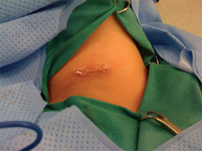
Our general approach is to make a 2.5-3.5 cm incision horizontally on the front of the neck. The size of the incision is determined by the patient’s neck size and other anatomic considerations. Through this incision we can access the position of any parathyroid gland in the neck and also monitor the function of the recurrent nerve. With the minimally invasive approach we dissect directly to the parathyroid gland ( adenoma) in question and remove it. Commonly, we identify the other parathyroid gland on the same side as the abnormal parathyroid gland. If the intra-operative PTH level drops appropriately ( > 50% within 15 minutes of parathyroid adenoma removal) and the other parathyroid gland is normal, the the chances of cure are greater than 95%.
The operative time is around 30 to 40 minutes. The patient is taken to the recovery room, and then observed for a few hours . If everything looks good, the patient may go home. There is very little recovery. The patient may return to work when they feel well, usually about 3-4 days. Most patients take only over the counter medication for pain. The wound is closed with skin glue and the patient may shower the next day.
The important features of minimally invasive parathyroidectomy are small incisions and minimal dissection. The main advantage is minimizing operative time. Minimal dissection ,in theory, reduces risk of injury to surrounding structures. That is to say, there is no un-necessary dissection or trauma to surrounding tissues. The approach described above is the most widely accepted among experienced parathyroid surgeons. Some surgeons use intra-operative sestamibi detection using a hand held probe to find the abnormal gland. The rest of the operation is similar to above, though a percent change in radioactivity is measured rather than the PTH level drop. The radio-guided approach is reported to be effective in experienced hands. We have had extensive experience with both techniques and have found the intra-operative PTH technique to be more reliable. Our cure rate with minimally invasive parathyroidectomy is about 95%. About 75% of patients are eligible for this approach. Importantly, the technology is a great advancement and very helpful. However, it is not perfect. As a result we have a very low threshold to examine all the parathyroid glands to ensure all abnormal parathyroid tissue is removed. With current techniques, a complete exploration may be accomplished through a very small incision. (View a minimally invasive parathyroidectomy performed by Dr. Faust)
Formal Neck Exploration
If pre-op imaging is negative then a complete neck exploration is performed. All 4 glands are identified and the abnormal gland is removed. The operation is done under general anesthesia. A 2.5-3.5cm incision is made depending on a variety of patient related factors. Operative time may be 1-2 hours. The wound is closed with skin glue. Most patients go home the same day. Recovery and pain issues are similar as with the minimally invasive approach. Again intra-operative PTH monitoring is done to document biochemical cure. Our cure rate is close to 98%. We use this approach when dealing with multi-gland disease (hyperplasia) and when pre-op imaging fails to identify an adenoma, or if there is any doubt that all abnormal parathyroid glands have been identified and removed.
The description above applies to 1°HPT due to a single adenoma. As mentioned earlier, about 15% of patients have multi-gland disease. Imagining may be confusing and even misleading with multi- gland disease. PTH monitoring may be helpful but is not perfectly reliable. This situation requires a great deal of experience to obtain optimal results. If all the glands are enlarged then 3 to 3 1/2 glands must be removed . One, or a portion of one parathyroid gland is left behind to function for the patient to prevent low calcium levels (hypocalcemia). If a double adenoma is present then both are removed and the other two glands are left in place. PTH monitoring is done and we look for a 90% drop in the PTH level to predict cure. Overall our cure rate with multi-gland disease is about 90-95%.
Parathyroid cancer is very rare, representing less than 1% of patients with 1°HPT. Generally patients are older and present with higher(sometimes very high) calcium levels, and PTH levels. The diagnosis is difficult to make preoperatively. Pathologic examination of the tissues may not be helpful. The best indication of cancer is gross invasion into local tissues. Complete removal of the tumor including any surrounding involved tissue is the best treatment. Lymph node dissection does not seem to help much. Unfortunately radiation and chemotherapy have little to offer.