Understandably, most people are anxious about the idea of laryngoscopy. However, with good local anesthesia and a very narrow flexible fiberoptic scope, the procedure is painless with very little local discomfort.
The first part of the procedure involves administering a local anesthetic and vasoconstrictor into the nose and the back of the mouth.
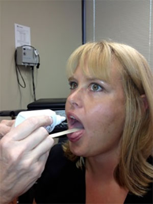
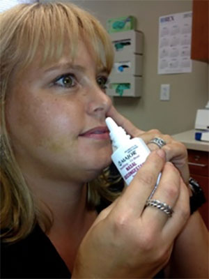
The patient then reclines for about 8-10 minutes. This allows time for the anesthetic to completely numb the nose and the back of the throat. The vasoconstrictor shrinks the mucosal lining of the nasal passages so the laryngoscope can pass easily through the nose to visualize the vocal cords.
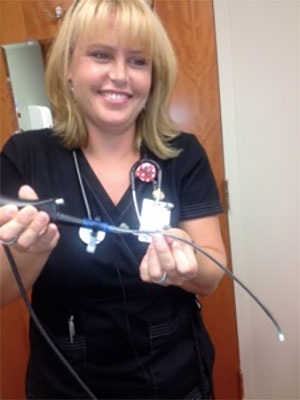
As you can see, the laryngoscope is quite small
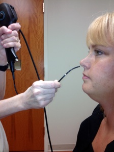
The initial part of the exam involves inserting the scope into the nose to the back of the throat. We curve the scope tip down so we can see the larynx, which is the upper part of the airway that contains the vocal cords.
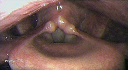
Once we see the larynx, we will ask you to Say “e” to close the vocal cords and assess their vibration. The picture below shows the larynx with the vocal cords closed.
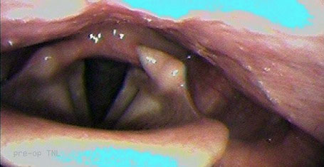
We will ask you to “sniff”. This opens the vocal cords, as shown below.
We also look for evidence of anatomic distortion, possibly due to the underlying thyroid pathology, or any other anatomic findings that could influence the approach to your operation.
The procedure lasts only a few minutes. Afterward, you are free to resume normal activity. You may drive, there is no sedation. However, it is important that you don’t eat or drink anything for about an hour after the procedure so that the feeling can return to the back of your throat. Oral intake before the anesthetic wears off could result in aspiration of fluid or food into your lungs.